

There are a variety of musculoskeletal issues that affect canine foot alignment. Some of these postural deviations are related to muscular weakness in the interosseous muscle and deep/superficial digital flexors, and can be improved with foot strengthening exercises. Others, however, require imaging and/or veterinary intervention due to damage to the tendinous or ligamentous structures.
First we will look at the qualities that define normal / correct canine foot alignment. Then we will identify the five most common postural deviations in the canine foot, examine the musculature affected by each. Lastly, we’ll discuss why some postural deviations can be improved with conditioning exercises, while others require veterinary intervention.
Normal / Correct Canine Foot Alignment
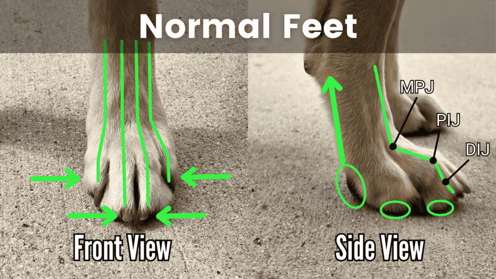
Correct foot alignment, viewed from the front, includes proper tension through the foot muscles, resulting in toes adducted / held tightly to the midline, with the proximal, intermediate, and distal phalanges (toe bones) properly stacked and supported.
Viewed from the side, the metacarpal/metatarsal pad (heel pad) is lifted, and the digital pads (toe pads) are fully grounded. We can also see the three joints of the toe properly aligned and supported with the MPJ, PIJ, and DIJ forming a step pattern.
- MPJ: Metatarsophalangeal Joint
- PIJ: Proximal Interphalangeal Joint
- DIJ: Distal Interphalangeal Joint
Understanding Normal Canine Foot Mechanics
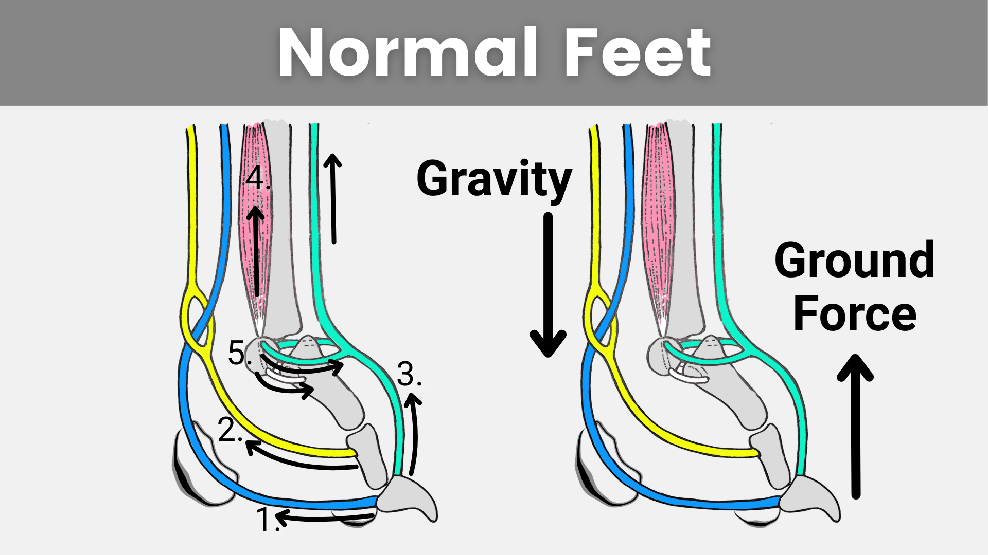
There are 5 main supporting structures that maintain the tensegrity of the canine foot anatomy. These include:
- Deep Digital Flexor (Blue): Runs along the back of the lower limb and inserts on the most distal phalanx (third toe bone / toenail). The primary action is to flex the toenails into the ground, and support the foot structure.
- Superficial Digital Flexor (Yellow): Runs along the back of the lower limb and forks around the deep flexor, inserting on the intermediate phalanx (second toe bone). The primary action is to flex the toes, and support the foot structure.
- Digital Extensor (Aqua): Runs along the front of the lower limb and inserts on the distal phalanx (third toe bone / toenail). Its primary action is to lift/extend the toes, and oppose the massive forces applied by the deep / superficial digital flexors.
- Interosseous Muscle (Pink): This often forgotten muscle runs along the back of the metacarpals/metatarsals and inserts into a small sesamoid bone, at the sesamoid complex. It is one of the only muscles located within the foot (the muscle fibers of the digital flexors are actually much more dorsal, with only the tendon extending into the foot itself), and serves as a suspensor mechanism to counteract gravity and ground reaction force.
- Sesamoid Complex (Multi): Including the Palmar/Plantar Sesamoid bones (grey), Collateral Sesamoid Ligaments (aqua), and Distal Sesamoid Ligament (white). These underappreciated structures oppose the pull of the digital flexors and interosseous muscle, and act like a “hub,” transferring forces from the toes to the lower limb, and vice versa.
When any part of this system is disrupted or out of balance, dysfunction follows, and the gravity and ground reaction force vectors shift. When impact forces are transmitted through the adjacent tissues, compensation and repetitive overuse injury are just a matter of time.
– – – – – – – – – – – – – – – – – – – – – –
Five Most Common Postural Deviations in the Canine Foot
Below we will cover how to visually assess for dysfunctional canine foot alignment so you can identify this dysfunction in your own dog. We will also unpack the underlying anatomy and physiology of each postural deviation so you can begin to understand how these misalignments impact your pup’s foot alignment in the short-term and long-term.
1. Flat Feet

In canines, flat feet or collapsed arches can be identified by three main alignment faults:
- Weight shifting backward onto the metatarsal/metacarpal pad.
- The MPJ (first toe joint) collapsing downward and being even with or lower than the PIJ (second toe joint).
- The distal phalanx (third toe bone) disengaging from the ground, with the cranial aspect (front) of the digital pad being off-weighted.
Anatomy of Flat Feet
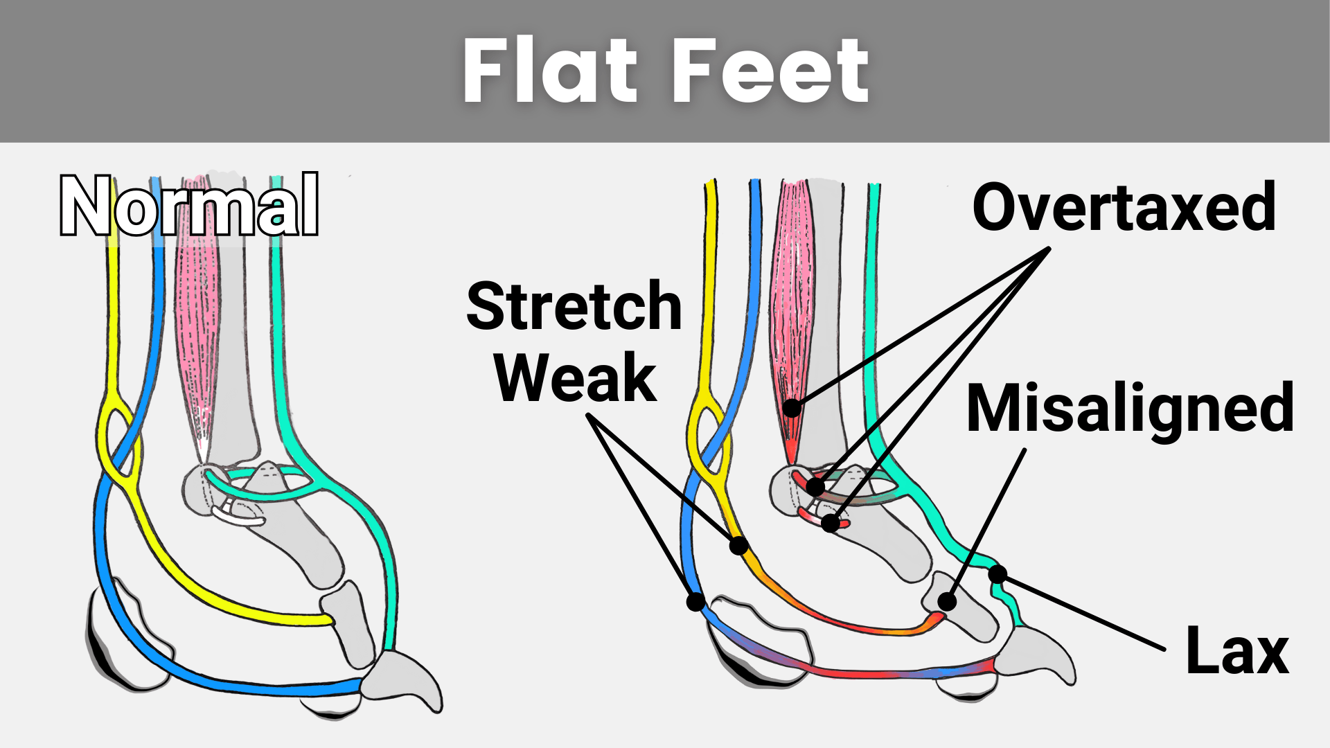
If we look at the anatomy of the foot, here we can see how the underlying anatomical structures are compromised:
- When a chronic backward weight shift occurs, the deep digital flexor (blue) and superficial digital flexor (yellow) are overloaded in a lengthened position and become stretch-weak as a result.
- This shifts load to the interosseous muscle (pink) and the collateral sesamoid ligaments, increasing the risk of sprain/strain-related injury to these structures.
- The digital extensor tendon (aqua) becomes lax as a result, increasing hypermobility and further compromising the alignment of the toe joints.
It should also be noted that left unaddressed, flat feet can progress into downed pasterns (front or rear), sprung toe / ruptured deep digital flexor and /or flat toe / superficial digital flexors, and even into plantigrade posture. It’s better to take steps to reconcile flat feet postural deviation now rather than taking a “wait and see” approach.
2. Rolling off the Toe Pads

Rolling off the Toe Pads (sometimes called Rolling onto the Heel Pad) is another very common postural deviation through the canine foot, and is characterized by the digital pads being “flipped up” and visible from the front and side view. Loss of contact between the digital pads and the ground also results in a backward weight shift onto the metacarpal/metatarsal pad.
Anatomy of Rolling off the Toe Pads
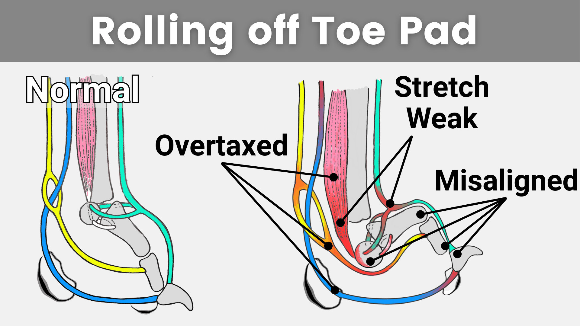
- Rolling off the Toe Pad Postural Deviation is the result of a weak interosseous muscle (pink).
- This weakness compromises the alignment of the sesamoid complex, causing the palmar/plantar sesamoid bone to drop downward and the phalanges to “kink.”
- The resulting poor alignment places the interosseous muscle and sesamoid collateral ligaments into a stretch-weak position (lengthened and chronically loaded).
- And overloads the superficial digital flexor (yellow) and distal aspect of the deep digital flexor (blue).
3. Splayed Feet/ Splay Foot
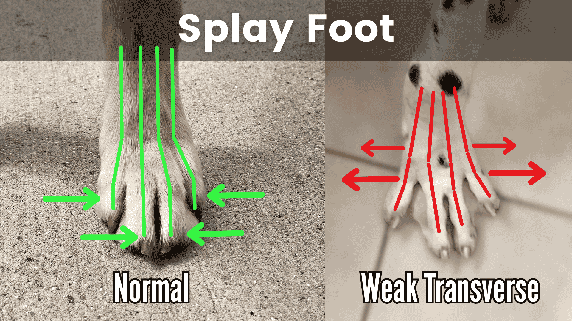
Splay Foot Postural Deviation is a misalignment on the dorsal plane and is characterized by the digits abducting out to the sides, revealing the interdigital webbing, coupled with a loss of support/collapse through the MPJ.
Anatomy of Splay Foot
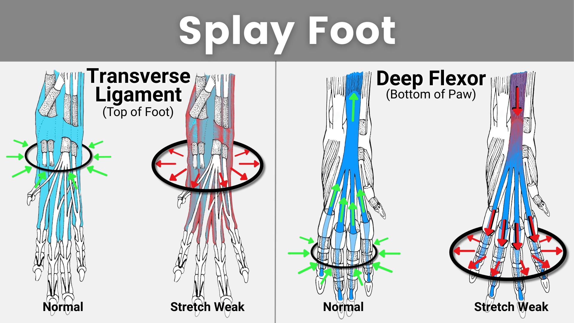
There are two main structures that prevent the foot from splaying, and support the metacarpals in the carpus/wrist and metatarsals in the hock from splaying:
- Transverse ligament (extensor retinaculum): This wraps around the carpus/tarsus horizontally, compressing the metacarpals/metatarsals inward and providing support for the digits… Just like a compression sleeve.
- Deep digital flexor: This has a secondary action of digit adduction (pulling the toes together) when in a tonic state (sustained contraction).
The transverse ligament (light blue) can be damaged by acute impact or by chronic/repeated stress. Alternatively, the transverse ligament can become lax due to systemic laxity which results from defective collagen fibers. Systemic laxity is being bred intentionally into some breeds as a means of producing loose skin and a lot of wrinkles, but all the connective tissue is affected, unfortunately. Additionally, Ehlers-Danlos (hEDS/hypermobility variety) has also been found in canines and can result in transverse ligament laxity and splay foot postural deviation.
Weakness, or lack of tonic engagement in the deep digital flexor (dark blue) can also contribute to splay foot postural deviation. When the deep digital flexor is lax/hypoactive and does not engage properly, the toes spread apart and lose functionality.
4. Sprung Toe / Damaged Deep Flexor Tendon

Sprung Toe is characterized by a distinct lifting of a single toe (or possibly multiples) in relation to the surrounding toes. The alignment of the remaining foot structure appears to be intact, but the second and third phalanx (toe bones) are lifted, with the digital pad losing contact with the ground.
Anatomy of Sprung Toe
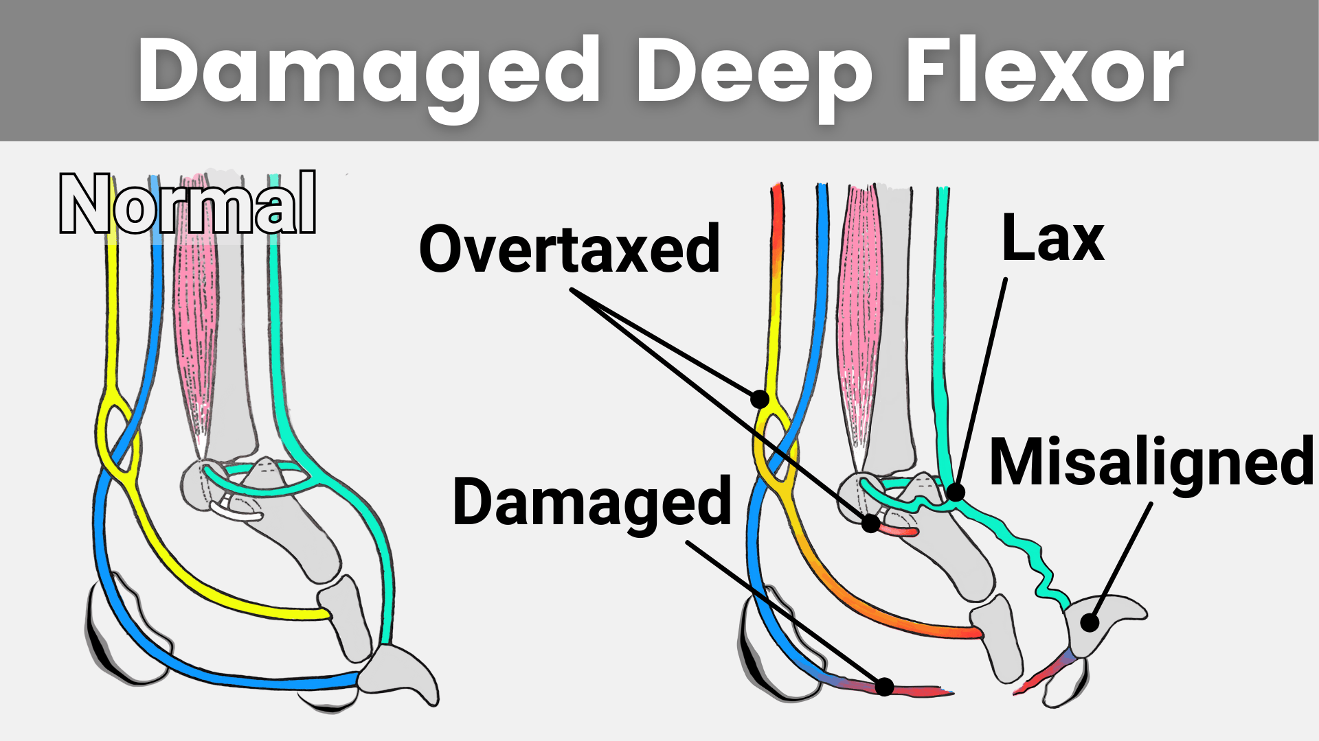
- Sprung Toe happens when the deep digital flexor tendon (blue) is damaged or ruptured completely.
- This leaves the third phalanx completely unsupported.
- The opposing digital extensor muscle (aqua) becomes lax, losing its ability to provide counterstabilization.
- This laxity is transmitted up the kinetic chain affecting the DIJ, PIJ, MPJ and Sesamoid Complex.
- The result is an overtaxing of the superficial digital flexor (yellow) and collateral sesamoid ligaments, as they become the primary shock-absorbing mechanisms in the canine foot.
While this is historically considered a *completely benign*… looking at how the underlying anatomy, and tensegrity interplay is compromised… I have a hard time believing that. This is especially true given that damage to the DDF in a human would always be addressed surgically to avoid long-term pain, and preserve foot mechanics.
5. Flat Toe / Damaged Superficial Digital Flexor

A Flat Toe is characterized by a single digit (or sometimes multiple digits) losing the stepwise alignment associated with normal/healthy foot alignment, appearing flat or overly long when compared to the surrounding toes. The digital pad itself maintains proper alignment, but the metacarpal/ metatarsal pad rolls under.
IMPORTANT: The images above are taken from the same dog. Please DO NOT confuse this postural deviation with a Hare Foot. Hare Foot conformation refers to the length of the first phalanx, and NOT the alignment of the phalanges, or muscle balance of the foot. No dog, regardless of breed, should demonstrate Flat Toe postural deviation. This is a hill I will die on.
Anatomy of Flat Toe
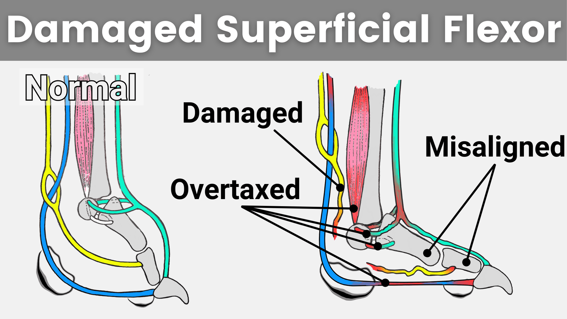
- Flat Toe happens when the superficial digital flexor tendon (yellow) is damaged or ruptured completely.
- This leaves the second and third phalanx unsupported.
- And overloads the deep digital flexor (blue), interosseous muscle (pink) and sesamoid complex / digital extensor (aqua) leaving them overtaxed, and more prone to injury in the future.
Similarly to Sprung toe postural deviation, this injury is historically considered benign. But, given the extensive alignment compromise to the canine foot’s structure, this assessment is questionable at best. Damage to the superficial digital flexor in a human would always result in immediate surgical intervention… Especially in an active human.
Dogs compensate well; however, humans are not always adept at accurately assessing pain in dogs, leaving many dogs to deal with chronic pain, and decreased quality of life.
– – – – – – – – – – – – – – – – – – – – – –
Can Conditioning Exercises Improve My Dog’s Foot Alignment?
Sometimes. If the postural deviation is related to muscle weakness, as is the case for Flat Feet, Rolling off the Toe Pad (collapsed arches), and Splay Foot, implementing a targeted exercise protocol can be helpful and very effective! Check out this post where I cover Four Foot Strengthening Exercises specifically designed to correct Flat Feet, Rolling off the Toe Pads, and Splayed Feet. They’re good for weak / downed pasterns as well!
When does Flat Feet require Veterinary Intervention?
Postural deviations that result from damage to tendinous or ligamentous structures often mean conditioning alone is not sufficient, and veterinary intervention is necessary. Since ‘sprung toe’ and ‘flat toe’ postural deviations are the result of damage to the flexor tendons, pursuing veterinary care is necessary.
Surgical Candidates
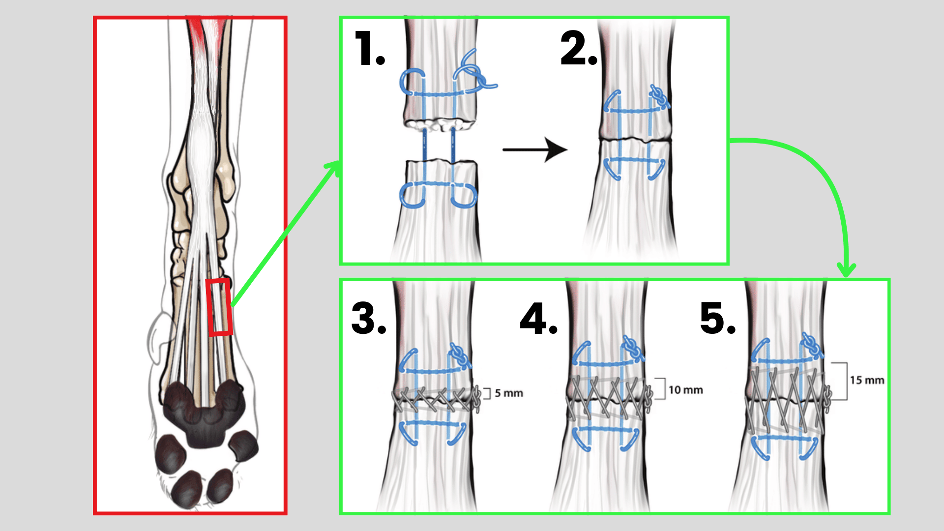
If you are reading this post and your dog has recently started to display this foot posture, immediate consultation with a board-certified orthopedic surgeon is necessary. Early diagnosis and intervention can significantly impact the long-term outcome and prevent further complications. In humans, primary surgical repair results in better functional outcome compared with secondary tendon repair (more than 3 weeks after the primary injury). After 3 weeks, primary tendon repair will not be possible because of proximal tendon end swelling, tendon contraction, and muscle fibrosis. While the specific protocols may differ in canines, the principle of timely intervention for tendon injuries remains critical.
Surgical repair of the deep/superficial digital flexor tendon should always be followed up by rehab to ensure proper mobilization of the surgical site and prevent entrapment in the flexor retinaculum. Consulting a board-certified sports medicine veterinarian or an experienced rehabilitation professional with CCRP or CCRT credentials is your best option for preventing post-operative complications.
After rehabilitation is complete, transitioning to canine conditioning long-term, to build strength through the digital flexors and surrounding musculature will be important to reduce the risk of re-injury and to re-train/un-train learned compensation mechanics that have likely become engrained during the recovery process.
Non-Surgical Candidates
If your pup has been demonstrating this postural deviation for a while (longer than 3 weeks), surgery is likely no longer an option. However, evaluation by a board-certified orthopedic specialist or sports medicine vet is indicated, and even if you have missed the surgical window or your dog is not a surgical candidate, there is still much that can and should be done!
Since we know joint hypermobility leads to an inflammatory response and bony spurring in humans, it’s not a stretch to assume the same would happen in canines. At the very least, a robust pharmaceutical pain management protocol should be integrated into the dog’s ongoing care routine, and the surrounding musculature should be strengthened to mitigate further joint misalignment. Lifestyle modifications should also be considered to limit / eliminate high impact activities given that the foot structure is now permanently compromised.
– – – – – – – – – – – – – – – – – – – – – –
Summary
Understanding canine foot alignment and the underlying compensatory mechanics is vital for both professionals and educated dog owners. By being able to visually assess for common postural deviations and recognizing when conditioning is appropriate versus when veterinary intervention is required, we can take proactive steps to support the health and overall well-being of our sport dogs and companion dogs.
Ultimately, training your eye to recognize these common postural deviations allows us to address issues early, prevent the progression of secondary conditions, and ensure our dogs remain functional and happy throughout their whole life.
0 Comments