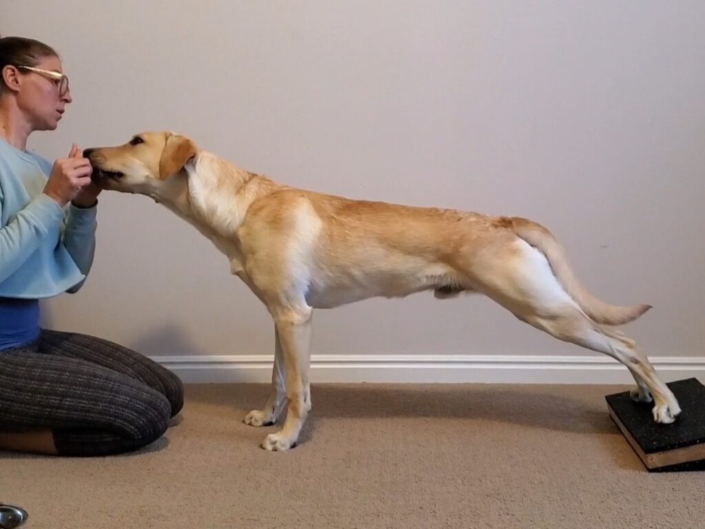
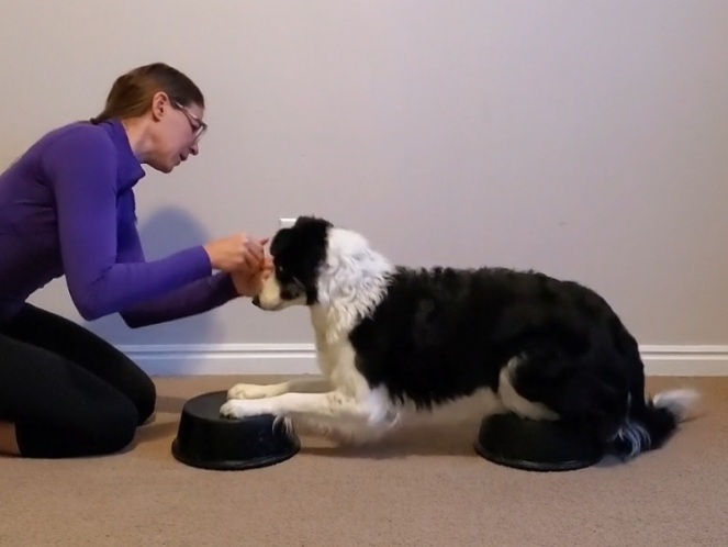
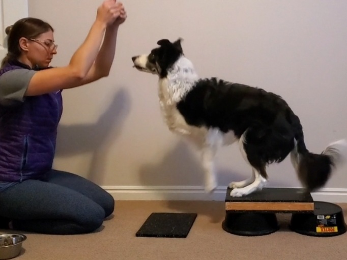
The Posture Sit exercise aims to improve your pup’s posture… and as a result targets primarily the postural muscles. These muscles are built for endurance, so they can hold the dog’s skeleton for a long time without fatiguing. For the most part, the muscles targeted in this exercise are working isometrically, which means without changing length or “without movement”. This Posture Sit Anatomy Breakdown explains how this musculature works together.
These muscles are split into 3 categories
Posture Sit: Muscles in the Core
These muscles affect and maintain the spine alignment. And because the Posture Sit exercise includes an alignment component, these muscles are trained both to move the spine into proper alignment (strength) and to hold it there (endurance).
- Epaxials: Intermediate to deep layer of muscles that run along the dorsal aspect of the dog’s spine. Extends the spine, and acts as a spine stabilizer. (Blue)
- Hypaxials: Deep layer of muscles that run along the ventral aspect of the dog’s spine, and includes the iliopsoas. Flexes the spine, and acts as a spine stabilizer isometrically. (Yellow)
- Iliopsoas: Compound muscle, comprised of the iliacus and psoas muscle. Part of the hypaxial group, and inserts on the proximal medial aspect of the femur. Flexes and/or stabilizes the spine, flexes the hip, and resists hip extension. (Teal)
- Internal Oblique: Intermediate abdominal muscle that lies deep to the external oblique. Flexes and/or stabilizes the spine if engaged bilaterally. Produces lateral spine flexion if engaged unilaterally (Lime green)
- External Oblique: Most superficial abdominal muscle that lies superficial the internal oblique. Flexes and/or stabilizes the spine if engaged bilaterally. Produces lateral spine flexion if engaged unilaterally (Yellow)
Posture Sit: Muscles in the Pelvic Limb
These muscles mainly affect the alignment of the pelvis and rear legs. The Posture Sit exercise, includes alignment that include having the stifle joint / femur pointing straight forward, and tracking parallel to each other. These muscles are responsible for internal / external rotation, as well as adduction / abduction.
- Adductor: Large inner thigh muscle. Adducts and stabilizes the femur, and stabilizes the pelvis. (Orange)
- Gluteals: Large superficial group of muscles running from the pelvis and sacrum to the proximolateral aspect of the femur. Abducts and/or stabilizes the femur. Extends and externally rotates the hip. If the pelvic limb is fixed, the gluteals will rotate the pelvis caudally. (Blue)
- Sartorius: Superficial muscle that lays on the cranial aspect of the pelvic limb, and runs from the iliac crest on the front of the pelvis to the medial aspect of the tibia. Because it crosses multiple joints it has many actions which include flexing and/or stabilizing the hip, cranial rotation of the pelvis and/or pelvic stabilization, and stifle extension. (Teal)
Posture Sit: Muscles in the Thoracic Limb
The Serratus Ventralis affects the alignment of the shoulder blade and ribcage. When engaged in the Posture Sit exercise, it helps the dog leverage the strength of the thoracic limb to help pull the thoracic spine (ribcage) into proper neutral alignment.
- Serratus Ventralis: Deep, fan shaped muscle that originates from the cervical vertebrae and ribs, and inserts on the medial aspect of the scapula. Stabilizes the scapula and assists in scapular rotation. The serratus has a “zig-zag” edge, similarly to that of a serrated knife. (Light green)
Feeling confused by some of this anatomy language? No worries! I made a Canine Anatomy: Glossary of Terms just for you!
